Multimodal Dataset of the Prostate
An exercise using 3D Slicer
Prostate Gland
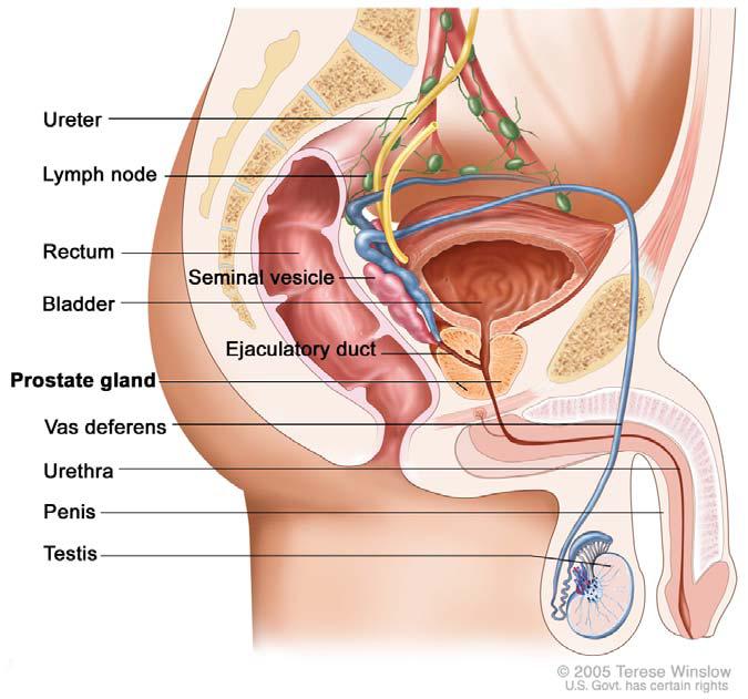
The prostate secretes the milky or white, slightly alkaline fluid, that constitutes roughly 30% of the volume of the semen.
A healthy human male prostate is classically said to be slightly larger than a walnut.

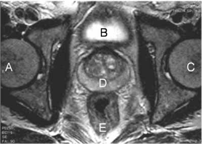
The prostate is located immediately in front of the rectum, a few centimeters from the anus. It surrounds the urethra just below the urinary bladder.
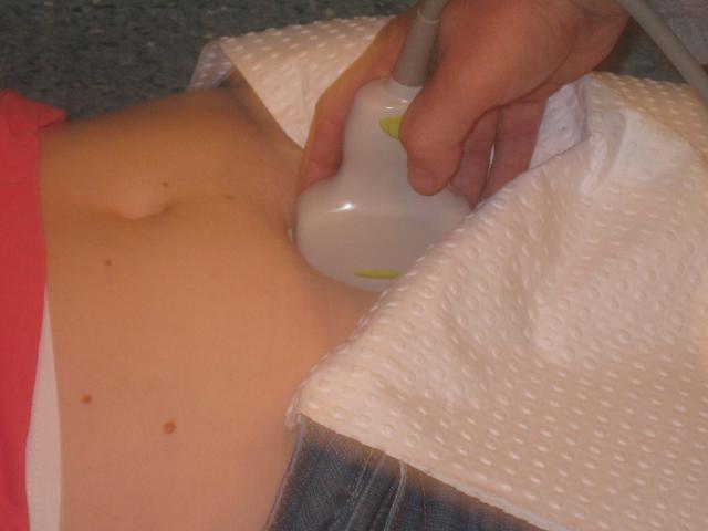
Ultrasound of the Prostate
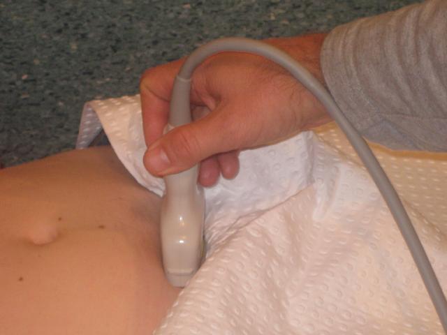
Transabdominal
In a transabdominal examination, the bladder is situated above the prostate
Sagittal
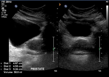
Transverse
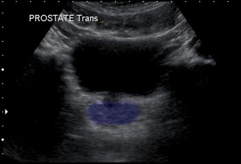
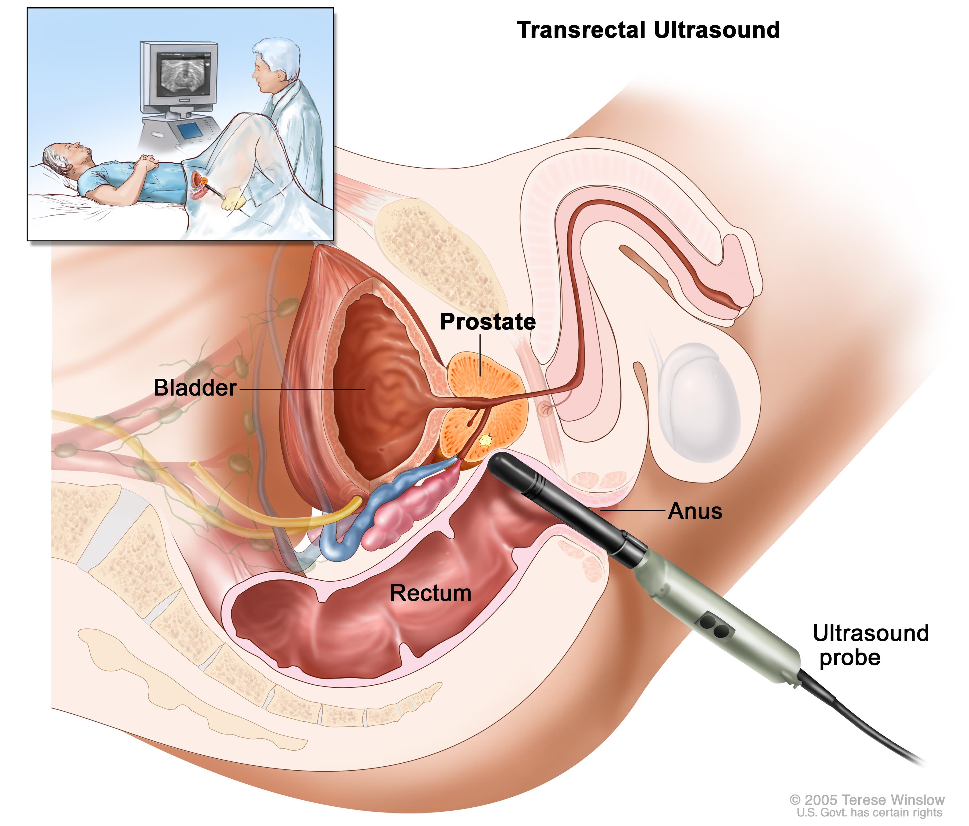
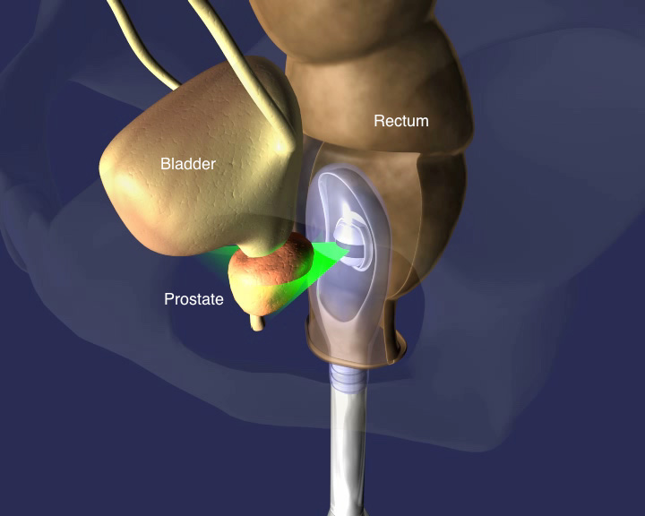
Transrectal
In a transrectal examination, the bladder is below the prostate (the transducer is now closer to the prostate than it is to the bladder)
Source: RadiologyWorld
Ultrasound images of the diseased prostate
MRI of Prostate

Slicer 3D
Load Sample Data
In the "Welcome to Slicer" screen, click on the "Download Sample Data" button.
Choose "Download MR-US Prostate"
Overlay volumes
MODULE: Set View Controllers Select "Four-Up" in the Windows View.

Choose the "View Controllers" Module in the module selector.
Use the following settings:

Volumes
Browse USProstate
- What are the dimensions of this volume?
- What are the dimensions of the voxels in this volume
- Change the LUT of the Ultrasound to "Magma"
- Where is the transducer positioned?
- Try to locate the prostate and the bladder
Browse MRprostate
- What are the dimensions of the pixels in this volume
- Change LUT to "WarmShade3"
- Set View Control transparency to 1.0 for MRprostate
- Find the Bladder. Hint: contrast has been added to the bladder
- Find the prostate. Hint, look for the crescent shaped structure as seen in the transverse view of the prostate in the MRI image above.
- Find the Urethra.
Volume Rendering
Select the Volume Rendering Module
-
Volume: MRProstate. Turn on eye
-
Display:
-
Crop: Enable. Display ROI
Crop the volume to the sagittal midline.
- What do you notice about the alignment of the MR volume to the axis
Slicer Scene
Slicer should now look something like this:

Create Segmentations
optional
Data Resolution
The resolution is very low in these volumes. You will not get the prettiest segmentations.
The prostate itself is too small and does not have enough contrast to segment using the "grow from seeds" tool. Instead, you would have to manually segment each slice of the prostate. We are not going to do that.
The Prostate appears to be smaller in the MR than in the US.
Bladder
- Bring up the "Segment Editor" module
-
For "Master Volume" - choose "MRProstate"
-
Add a segmentation for the bladder and call this segmentation "Bladder"
- Use Paint to paint inside bladder
- Add a segmentation for outside the bladder. Paint outside the bladder. Remember, you are just sampling voxels that are not bladder. You do not need a precision outline of the bladder
-
"Grow from seeds"
-
Select the Bladder segmentation
- Select "Initialize"
- Scrub through the slices and look for voxels with the wrong segmentation color
- Repair any wrong segmentations using the paint brush (be sure to select the right segmentation)
- Notice that you only need to repair a small segment and the tool will update the segmentation in that region
-
After you have checked all of the slices click "Apply"
-
"Show 3D" If you haven't done so, display the 3D view to see the result. You will probably see a large block
- Remove Outside segmentation
-
"Smoothing"
-
Use the default Kernel size
- Open
- Close
- Median
Rectum
Repeat the same process with the Rectum
IMPORTANT DIFFERENCES
-
"Grow from seeds"
-
Be sure to make the Bladder not visible (close eye next to segmentation)
-
Then, under the "Masking" tab, "Editable Area", select "Outside all visible segments"
-
"Smoothing" -

You should get something like this:
